ESCRTs Autophagy Coated vesicles Lysosomal regulation
The ESCRT System
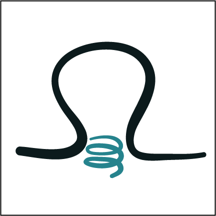
The ESCRT complexes catalyze one of the most unusual and mysterious membrane remodeling reactions in the cell. ESCRTs are responsible for the formation of multivesicular bodies (MVBs) in eukaryotic cells, remarkable structures that are required for transport of receptors and other membrane proteins to the lysosome. ESCRTs promote the budding of membrane away from cytosol, in other words, in the opposite direction from clathrin and other vesicle coats. The ESCRTs appear to promote this unusual mode of budding by forming an assembly at the bud neck, so as to temporarily stabilize the bud. The ESCRTs then go on to sever the membrane neck so as to release the bud. This membrane severing reaction is essential not only for MVB biogenesis, but also for the final severing of the narrow membrane neck separating two daughter cells during cytokinesis, for the release of microvesicles from the plasma membrane, membrane repair, the closure of autophagosomes, and for the release of HIV-1 and a number of other viruses from the plasma membrane of infected cells. Our group has been developing a structural hypothesis for this unique membrane budding and severing pathway. The current focus of the lab is to understand at the structural and mechanistic level how the ESCRTs are co-opted by HIV-1 so as to release nascent virions from infected cells.
Autophagy
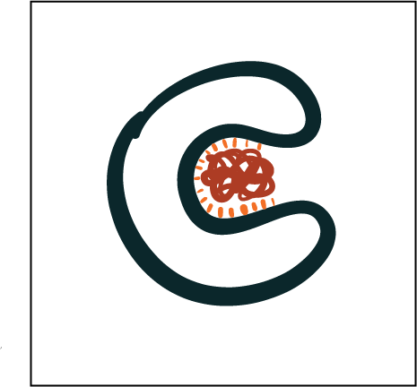
Autophagy is a form of cellular self-cannibalism that appears at first glance to be bizarre and harmful. Yet autophagy is conserved throughout eukaryotes and profoundly important for cellular health and survival. Defects in autophagy in single-celled organisms impair survival under starvation and stress. In humans, dysfunction in autophagy is linked to most of the major ailments of the developed world, including neurodegenerative diseases, cardiovascular disease, and cancer. Autophagy proceeds by the engulfment of cytosol, organelles, and inclusions by a growing double-membraned sheet. The double membrane becomes sealed and fuses with the lysosome (or vacuole in yeast), and its contents are hydrolyzed and recycled. Many, if not most, of the proteins required for autophagy have been identified, yet little is known about how they work with each other and with membranes to carry out this remarkable pathway. Our laboratory has solved the structures and worked out the interactions of many of the factors responsible for initiating autophagy. The group has had a strong focus on building up an integrated structural model for autophagosome formation in mammalian cells. We are now very focused on understanding the mechanisms of mitophagy (autophagy of mitochondria) in human cells because of its importance for understanding and treating Parkinson’s Disease.
Cargo sorting into coated vesicles
Host function and viral co-optation
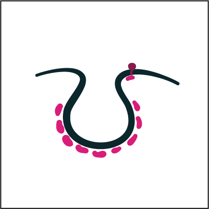
In comparison to the counter-intuitive and poorly understood ESCRT and autophagy pathways, far more is known about coated vesicle formation at the level of molecular mechanism. Because so much of the groundwork has already been done in this field, we can ask subtle and sophisticated questions about the structural mechanisms of regulation and the redirection of basic functions by pathogens. Cargo adaptors typically link membrane proteins to coat components. We have been focusing on the AP family of heterotetrameric adaptor complexes. We were recently able to obtain a structural explanation for how humans resisted infection by SIV, the ancestor of HIV, for millenia. We also recently began working on mechanisms of immune evasion by SARS-CoV-2, and determined the structure of its ORF8 protein, a fast evolving protein thought to be involved in downregulating MHC-I.
Lysosomal Regulation
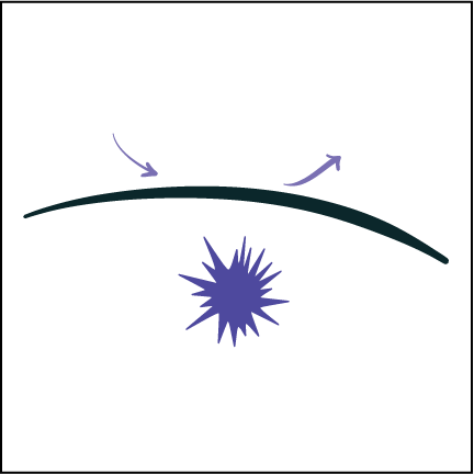
Lysosomes are not only the endpoint for degradative pathways, they are also a hub for signaling and sensing nutrient levels. With the Zoncu lab, we recently discovered the Lysosomal Folliculin Complex (LFC), which regulates the lysosomal small G-proteins RagA/B and RagC/D. We also found that the protein product of a major gene implicated in Frontotemporal Degeneration and ALS, C9orf72, participates in a complex analogous to the a portion of the LFC, and regulates the Arf family small G-proteins. These discoveries point to a rich and complex set of lysosomal signaling machines whose inner workings are attractive therapeutic targets in neurodegenerative diseases.